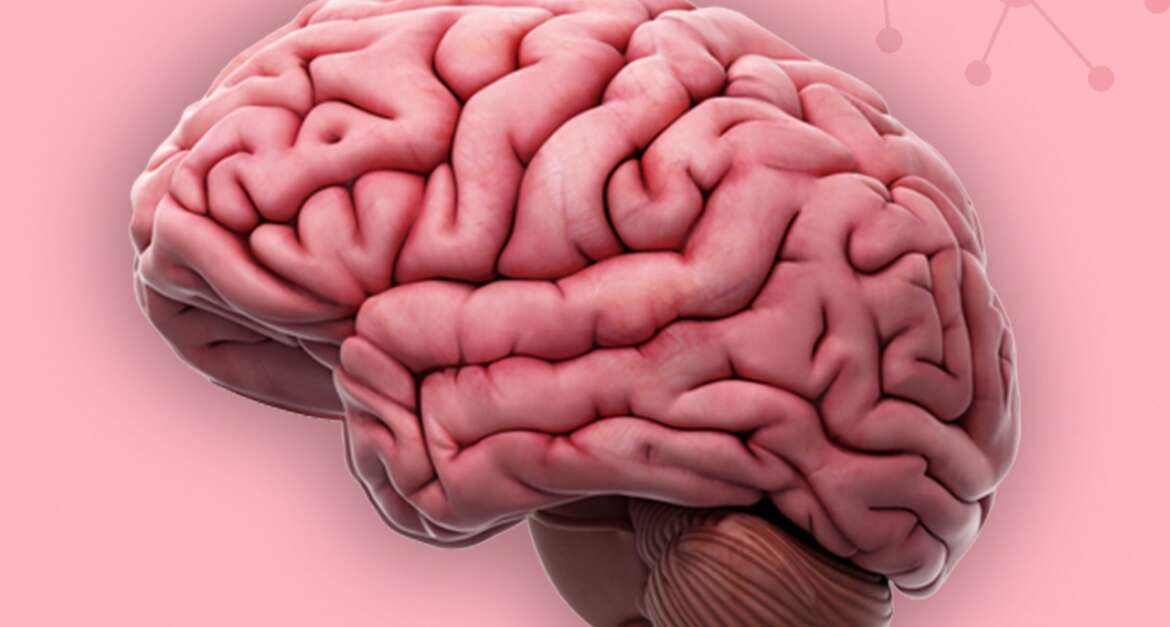
A condition is known as Chiari malformation, in which brain tissue extends into the spinal canal. The brain is forced downward when part of the skull is distorted or smaller than is typical.
Chiari malformations are rare, but the increasing use of imaging tests has led to a more frequent diagnosis.
There are three types of Chiari malformation, based on the anatomy of the brain tissue displaced into the spinal canal and whether developmental issues are present.
When the skull and brain are growing, Chiari malformation type 1 develops. Because of this, symptoms may not manifest until the late teenage or adult years. A congenital Chiari malformation such as type 3 or type 2 is present at birth.
Symptoms
Numerous people with Chiari malformations suffer from no symptoms and do not require treatment. Such people are only diagnosed through testing for unrelated conditions. However, the Chiari malformation can cause a variety of problems depending on its type and severity.
Chiari malformations are most commonly found in:
- Type 1
- Type 2
Signs and symptoms of these types are not as severe as those of the more rare pediatric form, type 3.
Causes
When the part of the skull containing the cerebellum (part of the brain) is too small or deformed, the pressure on the brain and crowding occur as a result of Chiari malformation type 1. There is an abnormal displacement of the lower portion of the cerebellum (tonsils) into the upper spinal canal.
Chiari malformation type 2 is always associated with a condition known as myelomeningocele.
It can disrupt the normal flow of cerebrospinal fluid, protecting the brain and spinal cord if the cerebellum is pushed into the upper spinal canal.
Impairment of cerebrospinal fluid circulation can block signals from the brain to the body or cause spinal fluid to build up in the brain and spinal cord.
The cerebellum may also press on the spinal cord or lower brainstem, causing neurological symptoms.
Factors associated with risk
Some families appear to be predisposed to Chiari malformations. Researchers are still investigating whether there is a hereditary component to the disorder.
Problems
Some people may develop Chiari malformation as a progressive disorder and experience serious complications. There may not be any associated symptoms for others, and no treatment is required. This condition may lead to the following complications:
- The Hydrocephalus. To relieve the accumulation of excess cerebrospinal fluid within the brain (hydrocephalus), a flexible tube (shunt) may need to be inserted to divert and drain the fluid.
- In the case of spina bifida. As a result of Chiari malformation, a condition known as spina bifida can develop. Complex situations such as paralysis can occur when part of the spinal cord is exposed. The Chiari malformation type 2 is usually associated with myelomeningocele, spina bifida.
- Syringomyelia. There is also an associated condition called syringomyelia, in which a cavity or cyst (syrinx) forms within the spinal column in people with Chiari malformation.
- Tethered cord syndrome. The spinal cord is attached to the spine and is stretched due to this condition. Nerves and muscles in the lower body may suffer severe damage as a result.
Diagnosis
A physician will examine you physically and review your medical history to diagnose your condition.
Additionally, your doctor may order imaging tests to diagnose your condition and determine its cause. Examples include:
- M.R.I. stands for magnetic resonance imaging.
- Safe and painless, this test provides detailed 3-dimensional images of structural differences in the brain that may be contributing to symptoms. As well as providing images of the cerebellum, it is capable of determining whether the cerebellum extends into the spinal canal.
- It is possible to repeat an M.R.I. over time and monitor the disorder’s progress.
- This is known as a computed tomography scan (C.T. scan). Other imaging tests, such as a C.T. scan, may be recommended by your physician.
- X-rays are used to create cross-sectional body images in a C.T. scan. A brain scan can reveal brain tumors, brain damage, bone and blood vessel problems, among other conditions.
Medications
Various treatments are available for Chiari malformations, depending on the severity and characteristics of your condition.
If you do not exhibit any symptoms, your doctor may recommend no treatment other than monitoring with regular examinations and magnetic resonance imaging scans.
You may be prescribed pain medication if headaches or other types of pain are your primary symptoms.
Reducing pressure through surgery
Symptomatic Chiari malformations are typically treated by surgery. It is intended to halt the progression of changes to the anatomy of the brain and spinal canal and relieve or stabilize symptoms.
Successful surgery can assist in reducing pressure on the cerebellum and spinal cord and restore the natural flow of spinal fluid.
During a procedure called posterior fossa decompression, a small section of bone is removed from behind the brain to relieve pressure on the brain.
The brain’s covering; the dura mater, may be removed in many cases. Additionally, a patch may be sewed into the covering to provide more room for the brain. Depending on the material, this patch might be artificial, or it may be made from tissue taken from another part of the body.
A small portion of your spinal column may also be removed to alleviate pressure on the spinal cord and provide more room for the spinal cord.
Surgical techniques may vary according to whether you have a fluid-filled cavity (syrinx) or if you have fluid in your brain (hydrocephalus). There may be a need for a tube (shunt) if you have a syrinx or hydrocephalus
Risks and follow-up after surgery
Infection, cerebrospinal fluid leakage, and wound healing problems are risks of surgery. Considering surgery as a treatment option, talk with your doctor about its pros and cons.
In most cases, the surgery will reduce symptoms, but it won’t reverse nerve damage in the spinal canal that has already occurred.
Your physician will need to perform periodic imaging tests, including regular follow-up examinations, following the surgery to assess the outcome and cerebrospinal fluid flow.

